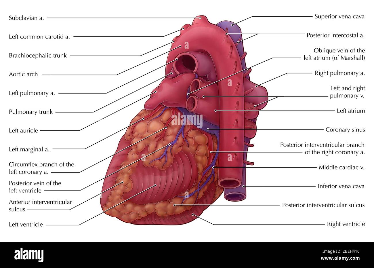
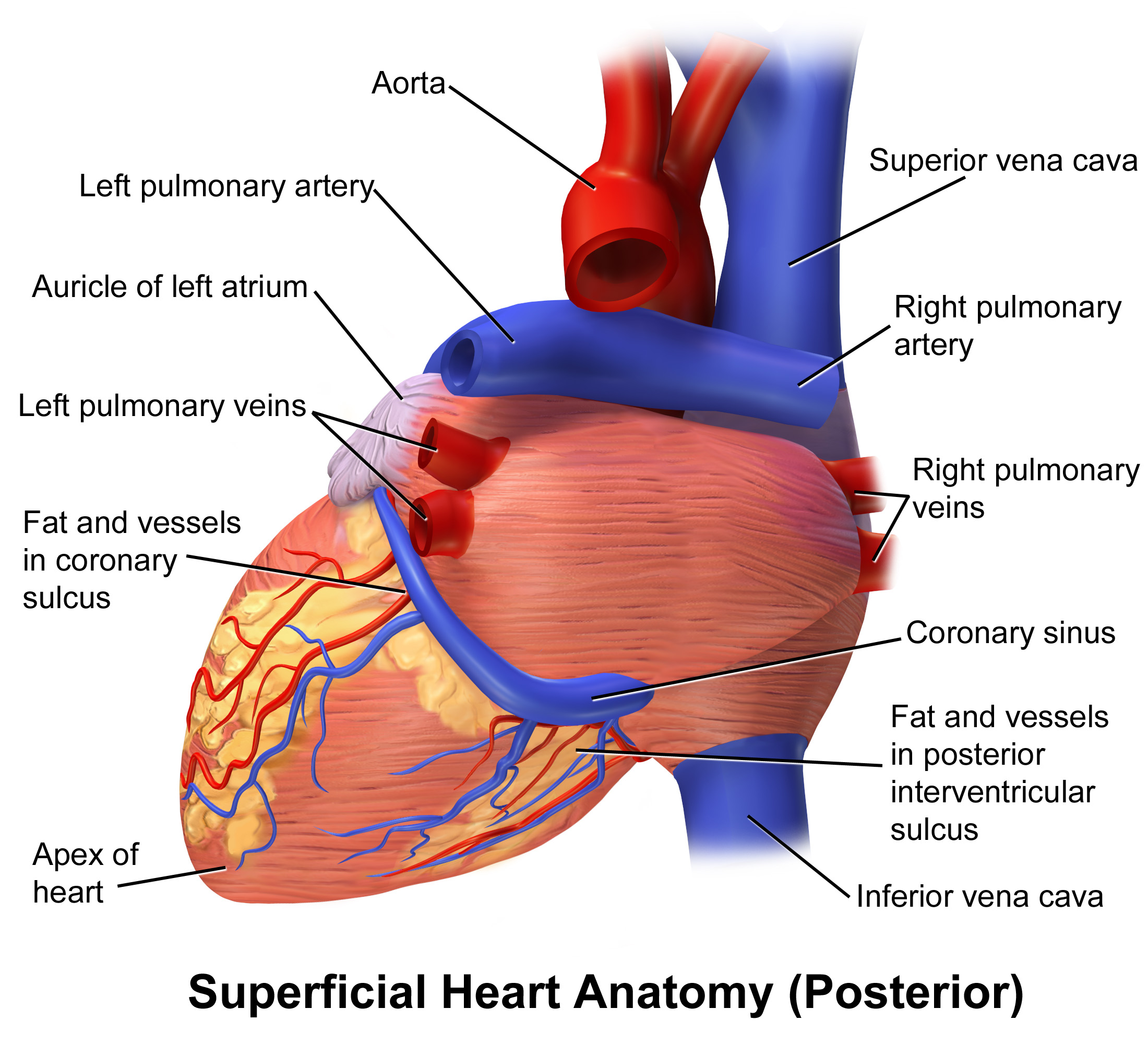
[最も選択された] heart anatomy posterior 102870-External heart anatomy posterior view
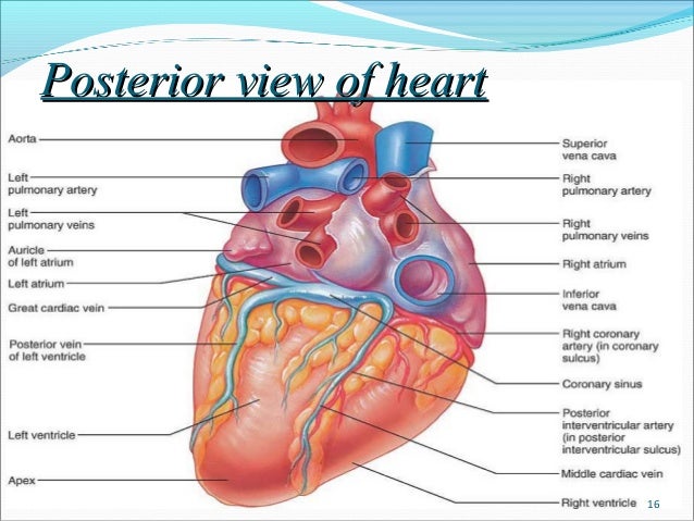
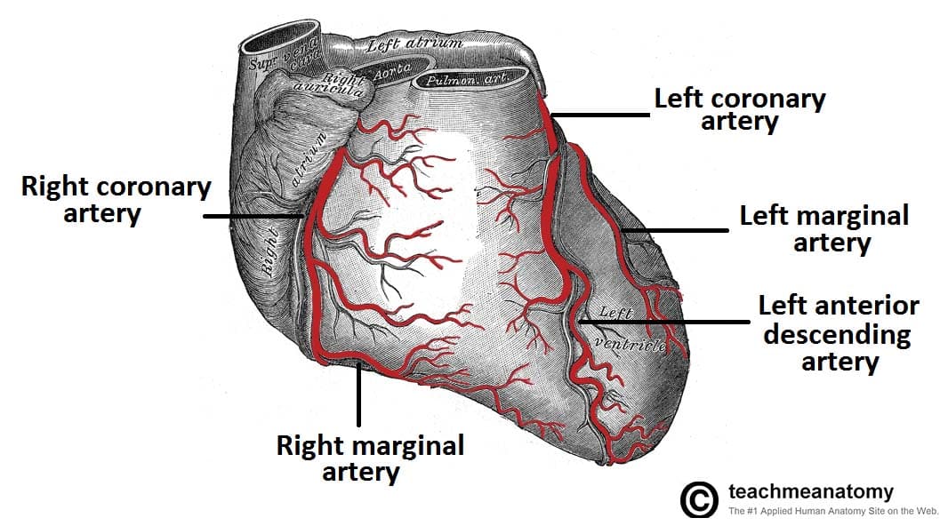
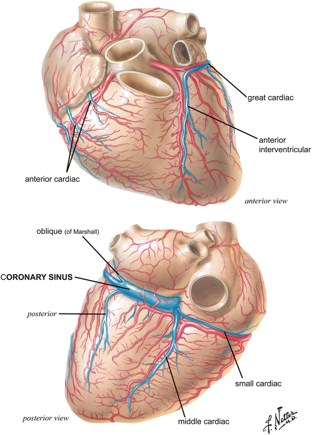
Venous Drainage of the HeartMost blood from the heart wall drains into the right atrium through the coronary sinus ,which lies in the posterior part of the atrioventricular groove It is a continuation of the great cardiac vein It opens into the right atrium to the left of the inferior vena cava30Jesús A Custodio MarroquínThe heart is located between the two lungs in the space referred to as the mediastinum (the space in the chest between the pleural sacs of the lungsRight coronary artery (RCA) The right coronary artery supplies blood to the right ventricle, the right atrium, and the SA (sinoatrial) and AV (atrioventricular) nodes, which regulate the heart rhythm The right coronary artery divides into smaller branches, including the right posterior descending artery and the acute marginal artery
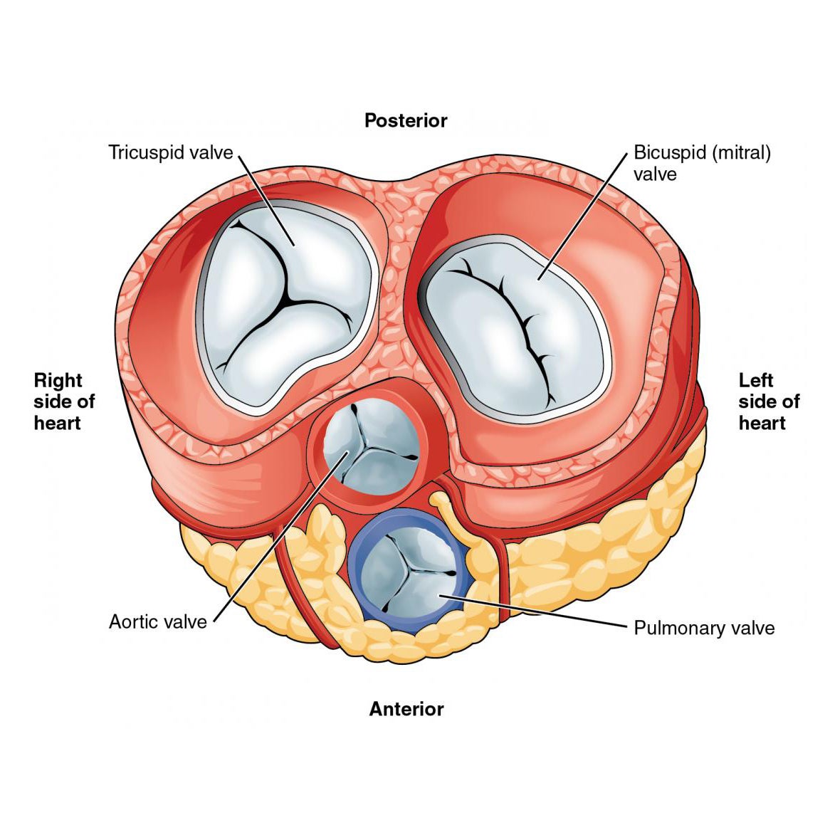
The Heart Anatomy How It Works And More
External heart anatomy posterior view
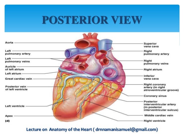
External heart anatomy posterior view-View Heartpptx from MCB 246 at University of Illinois, Urbana Champaign Week 2 Cardiovascular System Heart Anatomy and Physiology Lab II Figure 191 Components of the CardiovascularPOSITION Lies within the pericardium in middle mediastinum Behind the body of sternum and the 2nd to 6th costal cartilages In front of the 5th to 8th thoracic vertebrae A third of it lies to the right of median plane and 2/3 to the left Anterior to the vertebral column, posterior to the sternum Lecture on Anatomy of the Heart ( drnnamanisamuel@gmailcom)
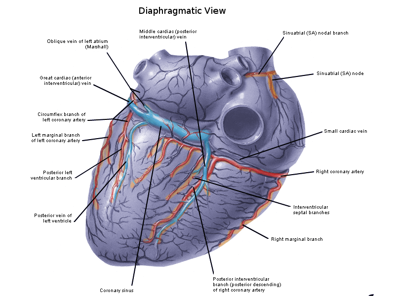


Anatomy Thorax Heart Veins Article
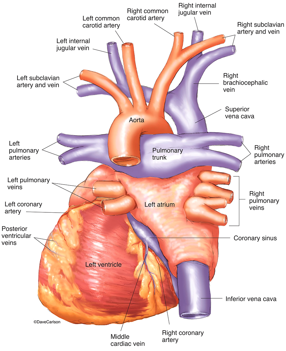
Posterior surface is now referred to as inferoposterior The posterior aspect of heart contains essentially the venous channels and the atrium (LA in particular)pulmonary veins and coronary sinus This happens right from 8 week heart open stage when venous end of lower straight heart tube folds up and posteriorlyWe are pleased to provide you with the picture named Heart Anatomy In Detail Anterior ViewWe hope this picture Heart Anatomy In Detail Anterior View can help you study and research for more anatomy content please follow us and visit our website wwwanatomynotecom Anatomynotecom found Heart Anatomy In Detail Anterior View from plenty of anatomical pictures on the internetLocation of the Heart The heart is roughly in a plane that runs from the right shoulder to the left nipple It lies in the protective thorax, posterior to the sternum and costal cartilages, and rests on the superior surface of the diaphragm;
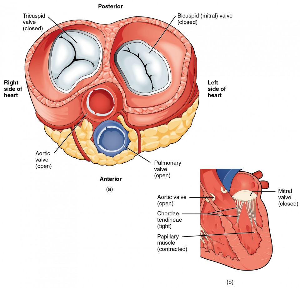
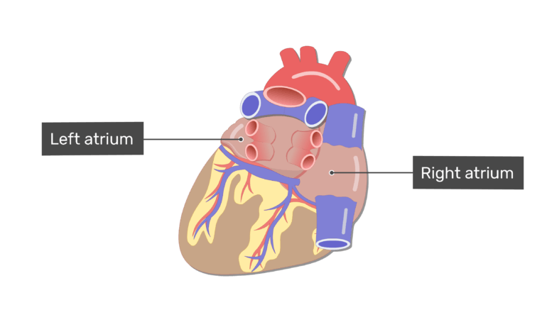
The left atrium is one of the four chambers of the heart, located on the left posterior side Its primary roles are to act as a holding chamber for READ MOREThe heart (Latin cor) is a thick, muscular organ with four cavitated parts located in the middle portion of the inferior mediastinumThe heart acts as a central pump of the cardiovascular system that maintains the unidirectional flow of blood The heart is a muscular organ with four cavities and it is located in the middle mediastinum, which is the middle part of the inferior mediastinumThere are two leaflets of flaps to the valve, one anterior and one posterior The valve is divided by imaginary lines into three anterior and three posterior segments The frame surrounding the valve to which the leaflets attach is called the annulus The leaflets are held anchored to the muscle of the heart through the papillary muscles
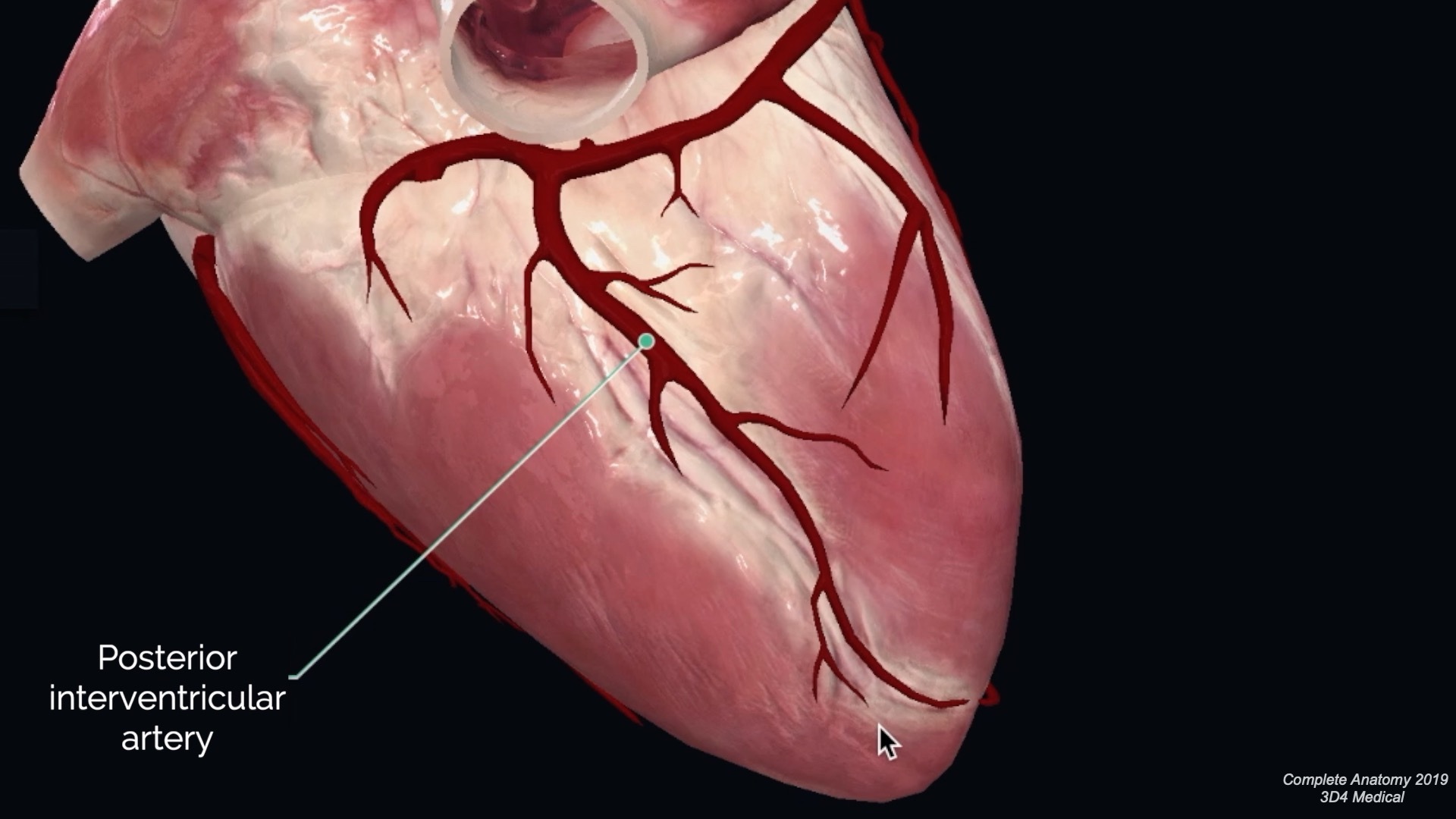
The heart is located in the thoracic cavity medial to the lungs and posterior to the sternum On its superior end, the base of the heart is attached to the aorta, Continue Scrolling To Read More BelowOn the posterior surface of the heart, the right coronary artery gives rise to the posterior interventricular artery, also known as the posterior descending artery It runs along the posterior portion of the interventricular sulcus toward the apex of the heart, giving rise to branches that supply the interventricular septum and portions of both ventriclesMost common signs of posterior circulation ischemia • Unilateral limb weakness (38%) • Gait ataxia (31%) • Unilateral limb ataxia (30%) • Dysarthria (28%) • Nystagmus (24%) Searls, DE, Pazdera, L, Korbel, E, Vysata, O & Caplan, LR (12) Symptoms and signs of posterior circulation ischemia in the New


Posterior Surface Of Heart
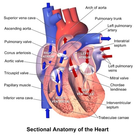


Right Ventricle Radiology Reference Article Radiopaedia Org
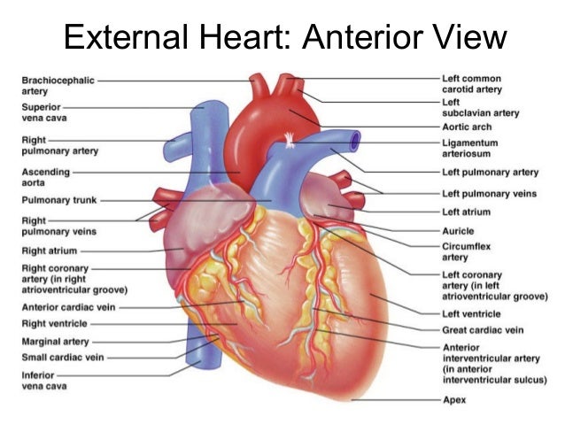
The anterior interventricular sulcus is visible on the anterior surface of the heart, whereas the posterior interventricular sulcus is visible on the posterior surface of the heart Figure \(\PageIndex{5}\) illustrates anterior and posterior views of the surface of the heart Figure \(\PageIndex{5}\) External Anatomy of the HeartPosterior side of the heart;Anatomy of heart 1 ANATOMY OF HEART Mr Binu Babu MBA, MSc (N) Asst Professor Mrs Jincy Ealias MSc (N) Lecturer 2 Heart • The heart is a hollow, coneshaped, muscular pump The heart beats about 25 billion times in an average lifetime
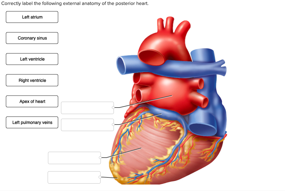


Solved Correctly Label The Following External Anatomy Of Chegg Com


Anterior And Posterior Views Of The Heart Showing Atheroma Royalty Free Cliparts Vectors And Stock Illustration Image
Anterior and Posterior Anterior refers to the 'front', and posterior refers to the 'back' Putting this in context, the heart is posterior to the sternum because it lies behind it Equally, the sternum is anterior to the heart because it lies in front of it Examples Pectoralis major lies anterior to pectoralis minorEmpties deoxygenated blood from the cardiac muscle into the right atrium Interventricular septum muscular wall that separates left and right ventriclesWebMD's Heart Anatomy Page provides a detailed image of the heart and provides information on heart conditions, tests, and treatments


Procedure B Dissection Of A Sheep Heart Human Anatomy Guws Medical
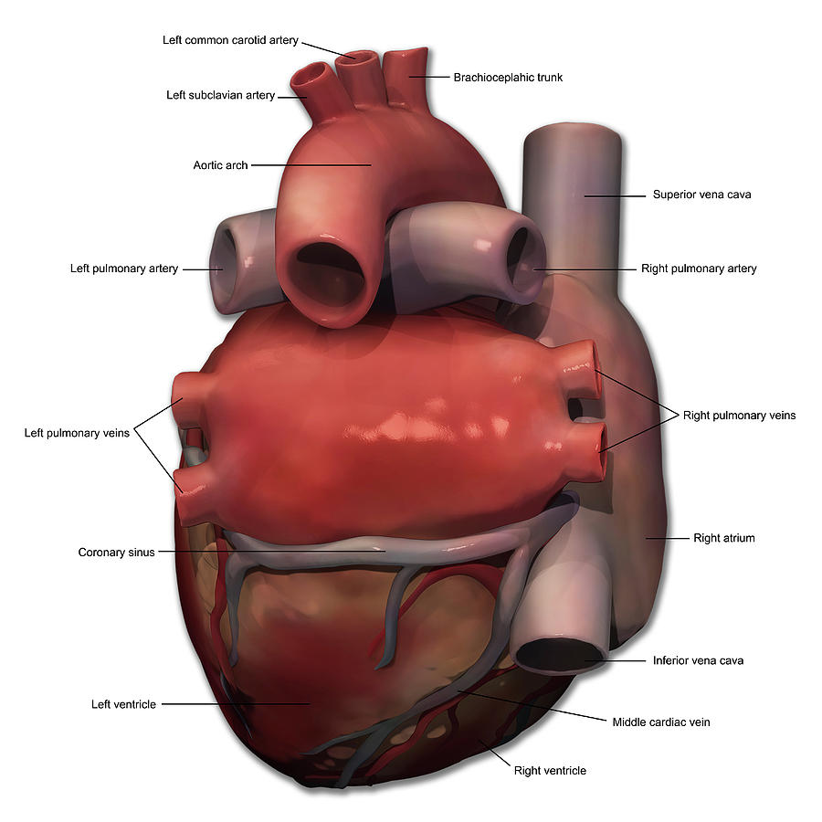


Posterior View Of Human Heart Anatomy Photograph By Alayna Guza
WebMD's Heart Anatomy Page provides a detailed image of the heart and provides information on heart conditions, tests, and treatmentsOn the posterior surface of the heart, the right coronary artery gives rise to the posterior interventricular artery, also known as the posterior descending artery It runs along the posterior portion of the interventricular sulcus toward the apex of the heart, giving rise to branches that supply the interventricular septum and portions of both ventriclesSUPPORT https//wwwgofundmecom/ninjanerdscienceNinja Nerds,Join us in this video where we show the anatomy of the heart in great detail through the use
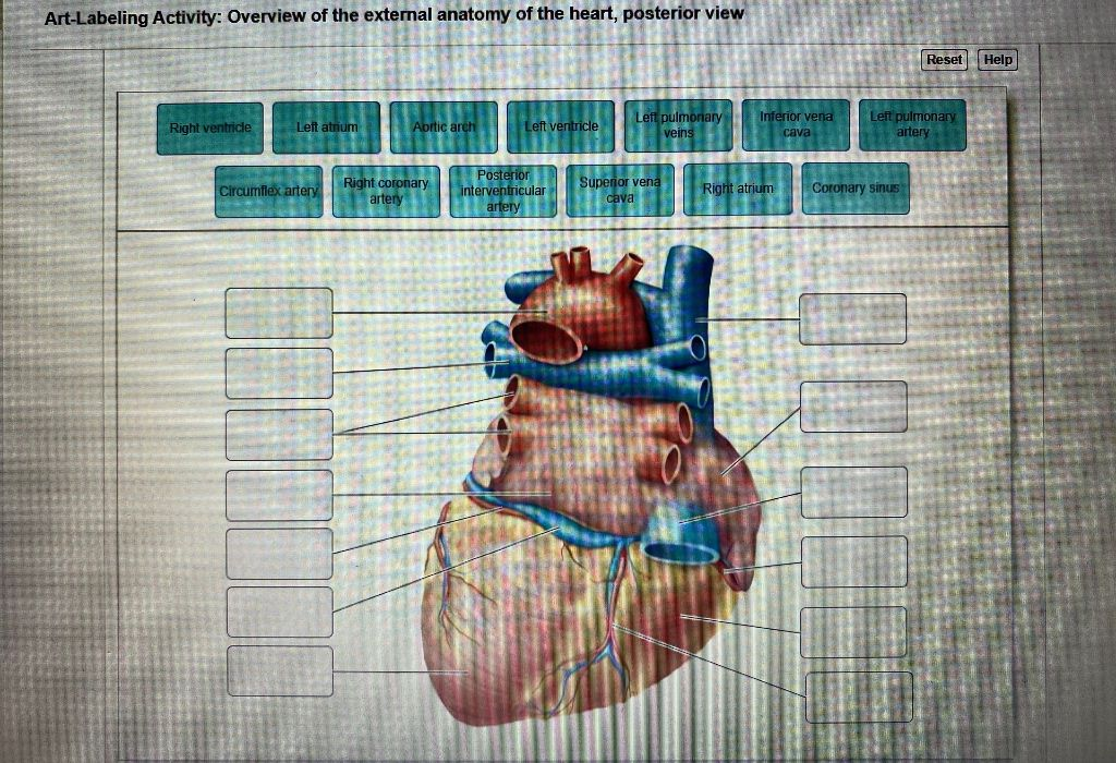


Solved Art Labeling Activity Overview Of The External An Chegg Com
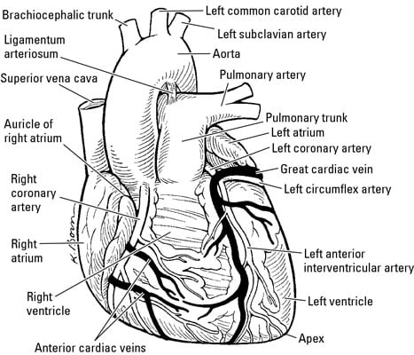


The Anatomy Of The Human Heart Dummies
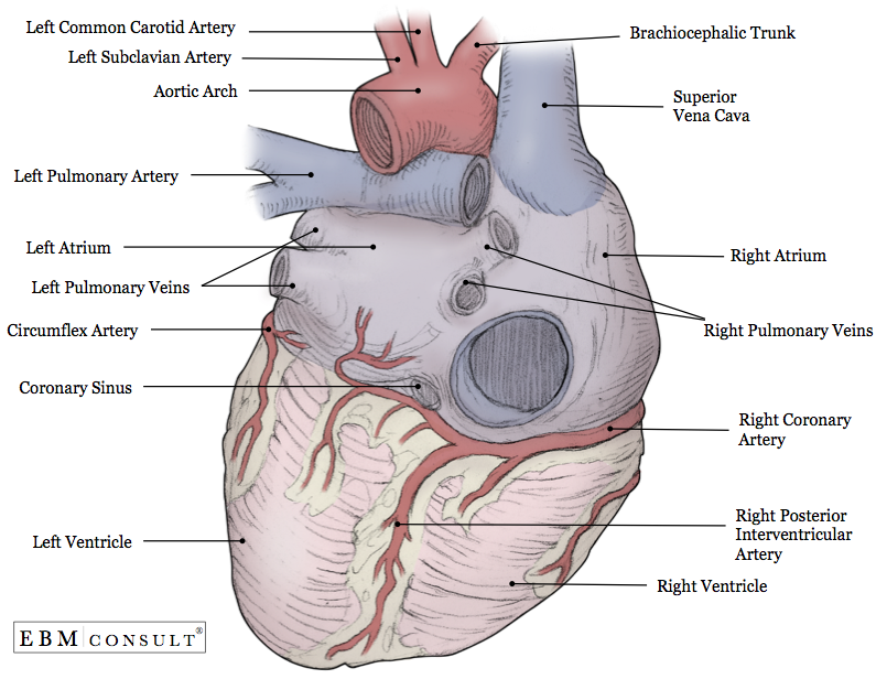
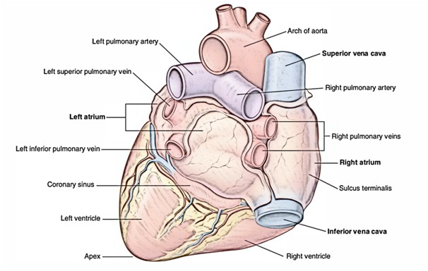
Heart anatomy this image shows the anatomy of the heart from external posterior view showing the different parts and features of the heart with the related vessels showing 1 superior vena cava 2 aortic arch 3 right pulmonary artery 4 left atrium 5 right pulmonary veins 6*Response times vary by subject and question complexity Median response time is 34 minutes and may be longer for new subjects Q Define resting membrane potential and describe its electrochemical basis A Neurons are a type of cells and a functional unit of the nervous system which is used toThe human heart is situated in the middle mediastinum, at the level of thoracic vertebrae T5T8A doublemembraned sac called the pericardium surrounds the heart and attaches to the mediastinum The back surface of the heart lies near the vertebral column, and the front surface sits behind the sternum and rib cartilages The upper part of the heart is the attachment point for several large
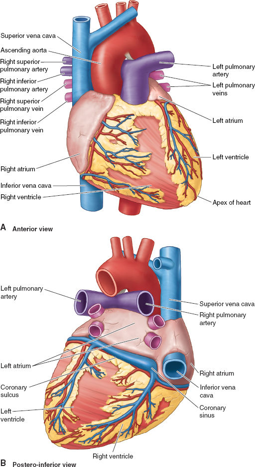


Cardiovascular Anatomy And Physiology Anesthesia Key
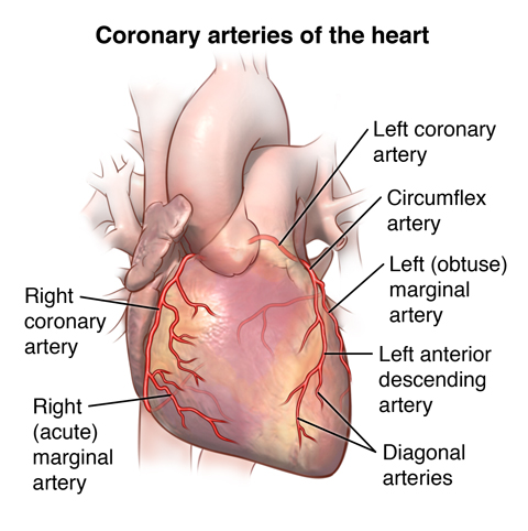


Anatomy And Function Of The Coronary Arteries Johns Hopkins Medicine
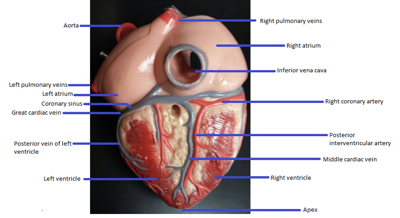
Heart Diagram Posterior view of the heart Saved by studentRN10 19 Heart Diagram Heart Anatomy Human Heart Cardiology Anatomy And Physiology Biology Medicine Animation WritingThe heart is a muscular organ about the size of a fist, located just behind and slightly left of the breastbone The heart pumps blood through the network of arteries and veins called thePosterior View Of Heart Anatomy In this image, you will find superior vena cava, right pulmonary artery, right pulmonary veins, right atrium, inferior vena cava, coronary sinus, right coronary artery, coronary sulcus, posterior interventricular artery in it You may also find posterior interventricular sulcus, middle cardiac vein, right ventricle, apex, apex of heart, left ventricle, posterior vein of left ventricle, great cardiac vein as well


The Heart Cardiovascular System


Pin On Anatomy And Physiology
Anatomy and Function of the Coronary Arteries The right coronary artery divides into smaller branches, including the right posterior descending artery and the acute marginal artery Together with the left anterior descending artery, the right coronary artery helps supply blood to the middle or septum of the heart This can lead to aThe posterior communicating artery is a potential location of aneurysms An aneurysm is a bulging area in an artery An aneurysm is a bulging area in an artery Although aneurysms in the circle of Willis most commonly occur in the anterior communicating artery, those in the posterior circulation account for 15% to % of all intracranial aneurysmsBecause the heart points to the left, about 2/3 of the heart's mass is found on the left side of the body and the other 1/3 is on the right Anatomy of the Heart Pericardium The heart sits within a fluidfilled cavity called the pericardial cavity The walls and lining of the pericardial cavity are a special membrane known as the pericardium


Human Heart Anatomy Posterior Page 1 Line 17qq Com


Http Eknygos Lsmuni Lt Springer 675 51 79 Pdf
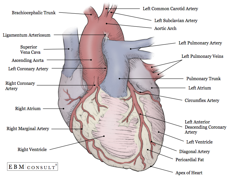
The heart has a somewhat conical form and is enclosed by the pericardium It is positioned posteriorly to the body of the sternum with onethird situated on the right and twothirds on the left of the midline Its leftsided orientation is formally known as levocardia (cf dextrocardia )Heart Heart anatomy The heart has five surfaces base (posterior), diaphragmatic (inferior), sternocostal (anterior), and Heart valves Heart valves separate atria from ventricles, and ventricles from great vessels The valves incorporate two Blood flow through the heart The blood flowIntraoperatively, the anatomy of the heart is viewed from the right side of the supine patient via a median sternotomy incision The structures initially seen from this perspective include the superior vena cava, right atrium, right ventricle, pulmonary artery, and aorta


External Anatomy Of Heart Posterior Part1 Diagram Quizlet


Posterior Interventricular Artery An Overview Sciencedirect Topics
The posterior interventricular artery, the ventricular branches of the RCA, the middle cardiac vein and the ventricular veins, all crossed the base of the space to their final destination AVNA originated from either the RCA itself or one of its branchesHeart Diagram Posterior view of the heart Saved by studentRN10 19 Heart Diagram Heart Anatomy Human Heart Cardiology Anatomy And Physiology Biology Medicine Animation WritingPosterior view of the heart H Hakeema Williams Heart Diagram Heart Anatomy Human Heart Cardiology Anatomy And Physiology Biology Medicine Animation Heart anatomy and physiology heart chambers and valves, heart vessels Where they're located, what they do and how they work Subclavian Artery


Heart Anatomy Class


View Of Human Heart Png Download Anatomical Heart Posterior View Transparent Png Vhv
The anterior interventricular sulcus is visible on the anterior surface of the heart, whereas the posterior interventricular sulcus is visible on the posterior surface of the heart Figure \(\PageIndex{5}\) illustrates anterior and posterior views of the surface of the heart Figure \(\PageIndex{5}\) External Anatomy of the HeartPosterior aspect of the heart Patients who are right coronary artery dominant have a branch off of the right coronary artery that supplies the posterior interventricular branch (or posterior descending artery) About 67% of patients are right dominant Function of the HeartThe posterior left ventricular (PLV) artery, also known as the posterolateral artery or branch (PLA or PLB), is a terminal branch of the coronary arterial system supplying the inferior portion of the heartIt usually arises from the right coronary artery in the typically rightdominant circulation as a terminal branch along with the posterior descending artery, but can less commonly arise from


Heart Anatomy Posterior Anatomy Drawing Diagram


Solved Rt Labeling Activity External Anatomy Of The Shee Chegg Com
Gross anatomy The heart has a somewhat conical form and is enclosed by the pericardium It is positioned posteriorly to the body of the sternum with onethird situated on the right and twothirds on the left of the midlineIt is located on the posterior surface of the heart The final 2 cardiac veins are also on the posterior surface of the heart On the left posterior side is the left marginal vein In the centre is the left posterior ventricular vein which runs along the posterior interventricular sulcus to join the coronary sinusThe heart, with its blood pumping function, is the very essence of life itself, which is why CPR must be commenced if a person's heart and breathing stops If you have a good knowledge of the human body, see how well you score in our heart anatomy quizzes


Amazon Com Heart Model Human Body Anatomy Replica Of Normal Heart For Doctors Office Educational Tool Gpi Anatomicals Industrial Scientific
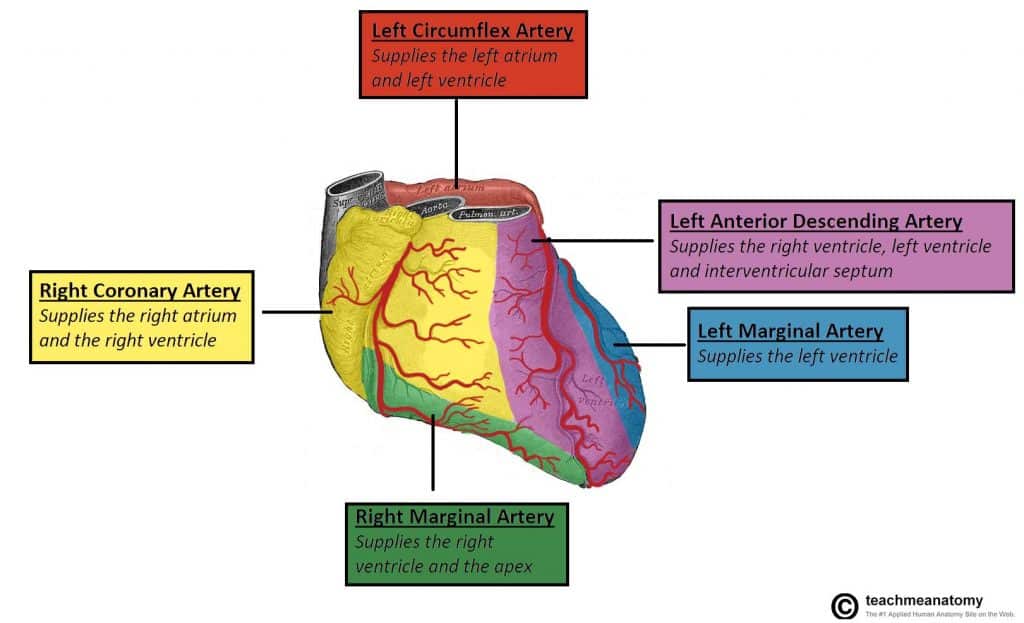


Vasculature Of The Heart Teachmeanatomy
General Heart Anatomy Posterior View Diagram Quizlet Anatomy of the human heart posterior view Heart anatomy posterior Putting this in context the heart is posterior to the sternum because it lies behind it The heart has five surfaces The heart is situated within the chest cavity and surrounded by a fluid filled sac called the pericardiumHeart Anatomy The heart is made up of four chambers Atria Upper two chambers of the heart Ventricles Lower two chambers of the heart Heart Wall The heart wall consists of three layers Epicardium The outer layer of the wall of the heart Myocardium The muscular middle layer of the wall of the heart Endocardium The inner layer of the heartIntroduction to Anatomy of the Heart This course is designed to give you a comprehensive introduction to the anatomy of the heart It is an interactive, lecture based course covering the underlying concepts and principles related to human gross anatomy of the heart and related structures


Your Heart Blood Vessels
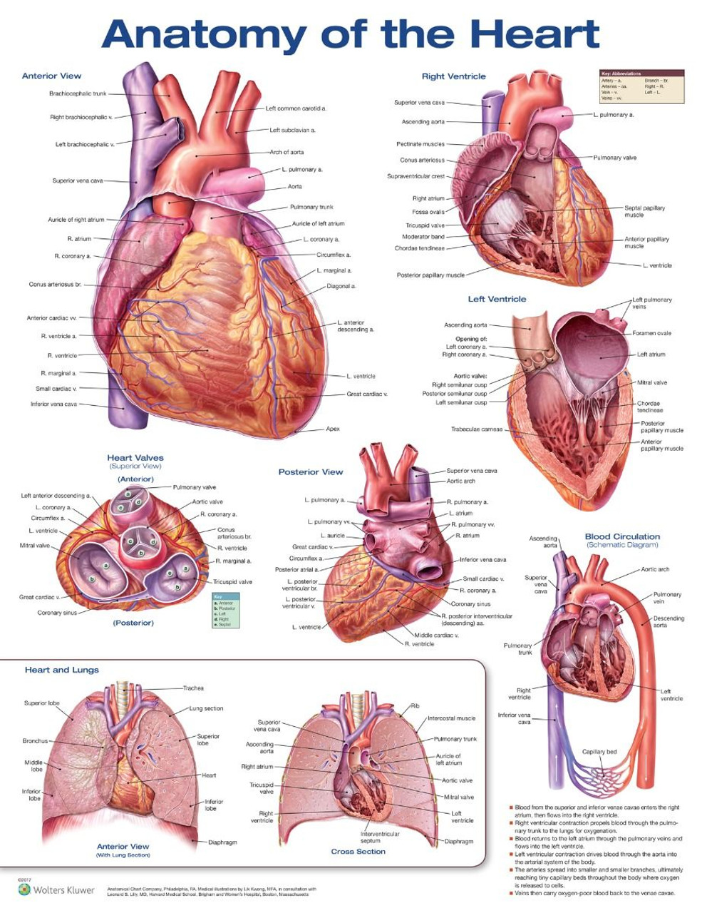


Anatomy Of The Heart 3rd Ed Clinical Charts And Supplies
Heart anatomy STUDY Flashcards Learn Write Spell Test PLAY Match Gravity Created by amontoya0519 PLUS Terms in this set (224) What part of the cardiac conduction system is located in the posterior wall of the right atrium, adjacent to the entrance of the superior vena cava SA node


Posterior Human Heart Anatomy Wall Decal Wallmonkeys Com


Anatomy Of The Heart And Major Coronary Vessels In Anterior Left And Download Scientific Diagram


Functional Anatomy Of The Cardiovascular System Clinical Gate
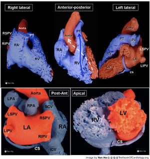


Anatomy Of The Heart Textbook Of Cardiology


Lungs And Heart Posterior View Illustration Stock Image C043 4863 Science Photo Library


Heart External Anatomy Posterior


Anatomy Of Heart
:background_color(FFFFFF):format(jpeg)/images/library/11110/Heart_Thumbnail.png)


Heart Anatomy Structure Valves Coronary Vessels Kenhub


Anatomy Of The Heart Surgery Oxford International Edition


Heart Anatomy Anatomy And Physiology Ii


Q Tbn And9gcr711 0ffebiiuw9gyabrsayorofjg Fjchbkqu4njezuy2m14r Usqp Cau


Anatomy Thorax Heart Veins Article


Anatomy Heart External


External Heart Anatomy Posterior Diagram Quizlet


Anatomy Heart External


Posterior View Of Heart High Resolution Stock Photography And Images Alamy


Heart Amboss


I Heart Anatomy I Heart Anatomy
/heart_electrical_system-597907ca03f4020010e78125.jpg)


Overview Of Sinoatrial And Atrioventricular Heart Nodes


The Heart Anatomy How It Works And More


Vasculature Of The Heart Teachmeanatomy


Coronary Arteries Wikipedia


Heart Artery And Vein Supplement Anterior View And Posterior View


Human Heart Posterior View Carlson Stock Art


Coronary Veins Cardiac Veins


Retired Human Heart Posterior Surface Anatomy Surfposh Jpg Human Heart Posterior Surface Anatomy Download
:background_color(FFFFFF):format(jpeg)/images/library/13372/n3ZFcGoK3SVFKEYB1gczCQ_R._interventricularis_posterior_01.png)


Posterior Interventricular Artery Anatomy And Supply Kenhub


3


Heart Anatomy Written Copy


Heart Models


Coronary System Tutorial What Is The Coronary System


Anatomy Of The Human Heart Springerlink


The Heart


Medivisuals Normal Heart Anatomy Medical Illustration


Chapter 5 Heart Dissection Anatomy And Physiology 2 Laboratory Manual


4 Posterior View Of The Human Heart Download Scientific Diagram


Pedi Cardiology Anatomy Coronary Veins Coronary Arteries Coronary Arteries Arteries And Veins Arteries Anatomy


Heart Anatomy Anatomy And Physiology Ii


Posterior Interventricular Artery Posterior Interventricular Human Heart Anatomy Human Anatomy And Physiology Heart Anatomy


Posterior Interventricular Artery An Overview Sciencedirect Topics


Heart Anatomy Anatomy And Physiology Ii


External Gross Anatomy Of The Heart Posterior View
:background_color(FFFFFF):format(jpeg)/images/article/en/heart/yh35Prra0VpHLwLZLrEDA_7dpDZkx6EDgihq981V55Qg_Apex_cordis_02-2.png)


Heart Anatomy Structure Valves Coronary Vessels Kenhub


Gross Anatomy Of The Heart Posterior View Diagram Quizlet


Coronary Venous Anatomy And Anomalies Journal Of Cardiovascular Computed Tomography


Heart Anatomy


Cardiac Anatomy Thoracic Key


What Exactly Constitute Posterior Wall Of Heart Dr S Venkatesan Md


Activity 1 Gross Anatomy Of The Human Heart And Using The Heart Model To Study Heart Anatomy Flashcards Easy Notecards


Heart Anatomy Class


Massasoit Instructure Com Courses Files Download Wrap 1


Coronary Circulation Wikipedia


Coronary Artery Anatomy Blood Supply To The Heart Geeky Medics


Anatomy Tutorial Posterior Atlas Of Human Cardiac Anatomy


Q Tbn And9gct Imnlh5wizmhmx18echk0zxb6jxawqbpcv0phtjegxwx5j0gz Usqp Cau


Posterior Interventricular Coronary Artery The Anatomy O Flickr


The Heart Chambers And Their Functions


Chapter 19 The Heart Circulatory System Heart Blood Vessels And Blood Cardiovascular System Heart Arteries Veins And Capillaries 2 Major Divisions Pulmonary Circuit Right


Cardiovascular Terms To Know
:max_bytes(150000):strip_icc()/heart_posterior_aorta-57f663543df78c690f0885c1.jpg)


Anatomy Of The Heart Aorta
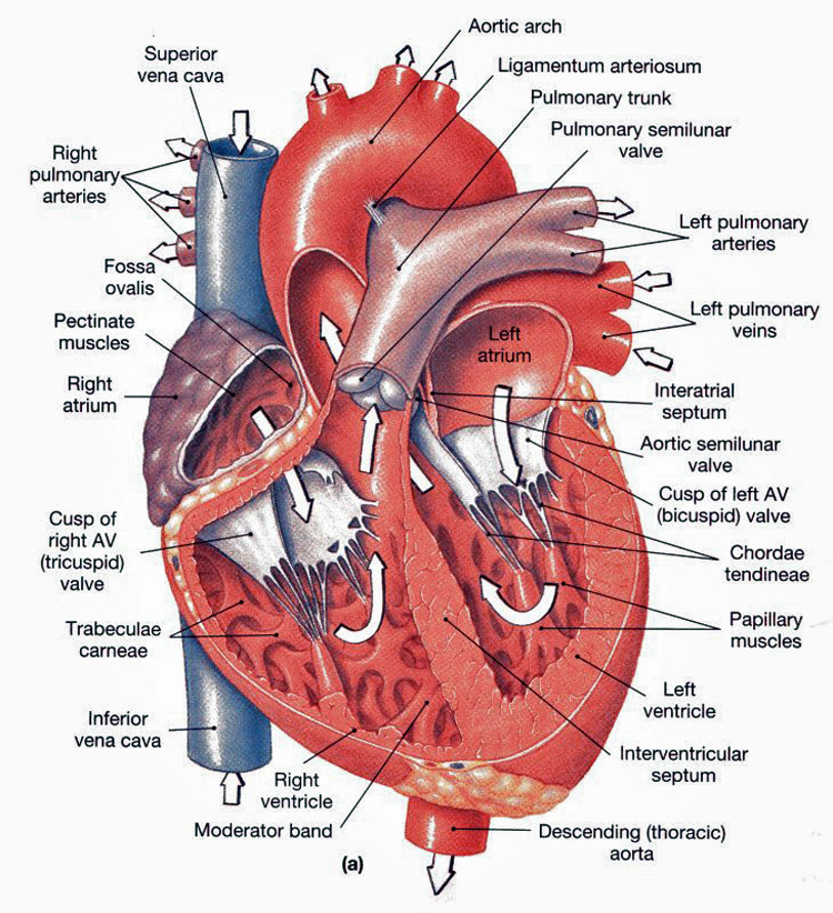


Heart Anatomy Chambers Valves And Vessels Anatomy Physiology


Study With A Pt Student Posterior View Of The Heart Ipad App


Q Tbn And9gcs4vuhnoufbevgxb6zoslqk V2sb8kvoeexlpglufaocnbfiuzs Usqp Cau


Posterior Heart Anatomy Anatomy Drawing Diagram


Cardiac System 1 Anatomy And Physiology Nursing Times


Heart Models


The Blood Vessels In The Heart Cardiovascular System
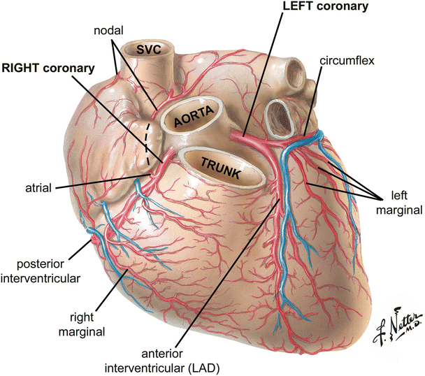


Anatomy Of The Human Heart Springerlink


Posterior View Of The Heart Heart Anatomy Heart Diagram Anatomy
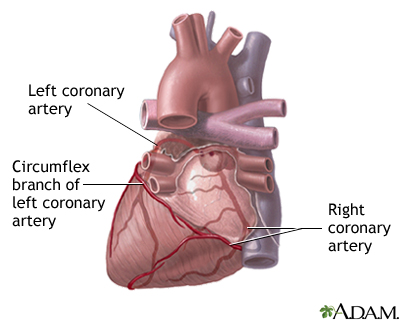


Posterior Heart Arteries Medlineplus Medical Encyclopedia Image


Human Heart Anterior Posterior Anatomy By Stanleyillustration On Deviantart


19 1 Heart Anatomy Anatomy Physiology


Human Heart Anatomy Isolated Stock Photo Download Image Now Istock


Cardiac Anatomy The Cleveland Clinic Cardiology Board Review 2ed


External Heart Anatomy Posterior View Diagram Quizlet


Coronary Sinus Cardiac Veins Anatomy Page 1 Line 17qq Com


Labeled Diagram Of The Heart Posterior View Anatomy And Physiology Heart Diagram Medical Coding


Edu Cdhb Health Nz Hospitals Services Atoz Publishingimages Pages Education And Development Module 1 anatomy and physiology of the heart 2 Pdf


File Blausen 0456 Heart Posterior Png Wikimedia Commons


Easy Notes On Heart Learn In Just 4 Minutes Earth S Lab


コメント
コメントを投稿